Case report. Lousy mind perfect brain. Early pseudodementia, frontotemporal dementia, nothing .2
Genetic Study
Negative for Psen1, Psen2, mapt & prgn.
Hippocampus Volumetric Study
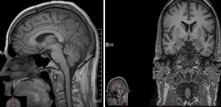
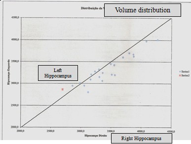
After a process of normalization and compared with normal healthy population for the hipocampal formations, the showed values are slight out of the defined normalized area, with a more reduced concentration in right hippocampus (see images below).
Left Sagital hemisphere & Frontal temporal coronal section

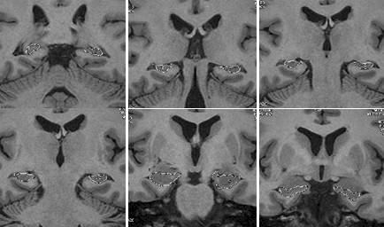
It is also in accordance with the idea that this relative process of atrophy of hypocampal formation affects mainly the respective bodies, with less significance in the hipocampal heads.
It is stated that in the valorization of these results, it has to be considered a slight bad proportion between the atrophic patterns of limbic formations, compared with the level of encephalic cortical atrophy (see images below).
Coronal sections of Hippocampus formation
(Volumetric sections from head & body – corpus callosum)

Graphic of volumetric measurements from bilateral Hippocampus formation

Electroencephalography
The results suggest etectrogenises constituted by Alpha activity with 10Hz frequency, medium amplitude, symmetric with predominance of posterior derivations. Activations by hyperventilation don not provoke pathologic graphic elements. Major Results: trace without signals of pathologic activity.
Magnetic Resonance results
Normal spaces sulci-cinstern and ventricular space in both intracranial compartments, in terms of permeability, configuration and dimensions. Focal asymmetries are not found. Regarding to signal presented, cerebral parenquima do not present considerable alterations. Some spaces of Virchow should be considered with no relevance.
A more detailed study, focused on hipocampal formations and rest of temporal lobes shows a reduced volumetric structure, mostly in the body of hippocampus and particularly at the right hemisphere. It is also observed a minor hiposignal in the Fast Flair Sequence (T2), at hippocampus level.
PET SCAN
In the PET Scan Study we can see a generalized cortical reduction on captation of radiopharmac, partially preserved in visual occipital cortex and in basal ganglia as well as in thalamic nucleus. We do not found regional deficits suggestive of Alzheimer Disease or Frontal Temporal Dementia.
Although the pattern found of cortical hiphometabolism, with preservation of visual cortical cortex are not typical for AD or FTD, degenerative microangiopathic and / or encephalitic pathology could not be excluded.
General clinical conclusions
The patient presented some deviant values in some clinical parameters but nothing that could justify the dramatic clinical picture. The only clinical examination not pursed was a cerebral biopsy due the very intrusive nature of this procedure.
The patient is undoubtedly disorganized in his capacity to cope with daily life responsibilities (not only socially but familiarly speaking). His ability to work was profoundly reduced, and a neuropsychiatric frontal-temporal syndrome is unquestionably present (despite the unknown ethiopatogeny).
This intriguing clinical case must, by that, be strongly discussed.
Discussion
One of the first hypotheses in the comprehension of this case is to consider if we were dealing with an unusual case of early pseudo dementia. Let us look at it.
The term pseudo dementia was first utilized by Wernicke, related with cases that comprised chronically hysterical states, as stressed by Bleuler (1934, in Bulbena & Berrios, 1986).
Nussbaum (1994) states that this term, “ pseudodementia” has remained a permanent nosological entity in the literature for over 100 years. He states that the identification of the fact that clinical symptoms related with reversible neuropsychiatric circumstances could imitate permanent disorders is accepted since the middle of the 19th century. As the author states: “The importance of the term lies in the inherent assumption that the presenting dementia is not real, or is at least reversible, and therefore treatable. Nonetheless, there continues to be controversy regarding the validity and appropriate clinical use of the term” (p.71).
Jones, Tranel, Benton & Paulsen (1992) made neurological evaluations in 37 patients (older than 45 years) showing early and subtle alterations changes in memory, in order to explore the efficacy of several neuropsychological tests in the distinction among dementia and pseudo - dementia. With a six months interval of “test” “re – test” the authors found major relations between faulty temporal orientation, visuo construction, and visual memory at Time 1, and the subsequent development of dementia.