Ovarian fibroid in pregnancy. Case presentation.2
Operation findings
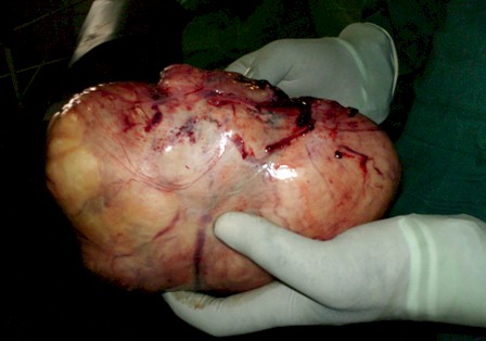
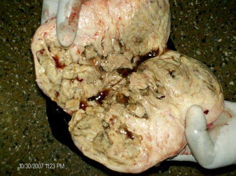
Normal uterine size for immediate post delivery, right tube and ovary were normal. Left ovary grossly increased in size measuring 20cm x 12cm, solid with smooth and regular surface. Left anexectomy was done. Hemostasis secured. No ascitis, no other lesions seen on omentum or liver surface. The cut surface on the tumor was whorled with trabecular appearance. No evidence of hemorrhage or necrosis. Biopsy specimen from omentum was also taken.
Patient progressed successfully and was discharged after six (6) days. She was seen again after a month when biopsy report was available. The biopsy report was consistent with ovarian fibroid.
Discussion
Ovarian fibroid is one of the less frequent tumors and its association with an uneventful pregnancy is extreme rarity. This tumor is not common in the adolescent age. There are several reports about higher incidence in the post-menopausal age but some studies have not found any difference between pre and post menopausal women as far as incidence is concerned. In nine years studies, Wai Leung and Pong Mo Yuen of Prince of Wales Hospital, University of Hong Kong, China, found that out of twenty three (23) patients who were diagnosed of ovarian fibroid, representing just 1% of all benign ovarian tumors, 47.8% were in post menopausal age and the rest were premenopausal women.
The average age of these patients was 45 years. In this particular case that we presented, the patient was in the reproductive age and tumor co-existed with an uneventful pregnancy until labour. The average size of these tumors according to some publications is 13cm and generally unilateral. Our patient had unilateral left ovarian tumor that measured 20cmx12cmx8cm.
This condition is generally asymptomatic until it acquires large volume where pressure symptoms may ensue. This is due to compression on pelvic and abdominal organs. Its diagnosis is generally accidental during pelvic ultrasound scanning or in laparotomy.
On ultrasound, ovarian fibroma appears as a hypoechoic mass with attenuation of the ultrasound beam. On MR, these masses demonstrate low signal on T2 relative to the myometrium due to the predominantly fibrous composition of the mass. On CT, ovarian fibroma is a well-defined, solid mass with mild heterogeneity. Ovarian fibroma may calcify and/or exhibit cystic degeneration. They are bilateral in 3-10% of cases. Ascitis is present in 10-15% of cases, especially with larger lesions.
Because this is a benign tumor, anexectomy is the standard treatment. Dr. Herbert R. Spencer report two cases that were successfully managed with anexectomy. In their publication in the Gynaecologist investigation journal, Wai Leung and Pong Mo Yuen also recommend anexectomy as the choice of treatment since these are benign tumors. In the case we presented, left anexectomy was successfully performed.


Bibliography
1- González - Merlo. J. Ginecología. 5ta. edición. Barcelona: Salvat editors, 1988: 520-523.
2- Penaloza A, Rodriguez D, Spinetti G, Pepe G, Lorenzo F: Tecoma, Presentacion de un caso clinico. Revision de la literatura; Gac. Med Caracas 1995, 103 (4):375-378.
3- Herbert R. Spencer M.D: Two cases of ovarian fibroid complicating pregnancy; pudmedcentral.nih-gov/articlerender.fcgi?artid=2046659
4-Wai Leung and Pong Mo Yuen: Ovarian Fibroid: a review on the clinical characteristics;karger.com/produkte.asp?typ=fulltext&file=G01200602001001.
5- Pelusi G, Taroni B, and Flamigni C. Benign ovarian tumor, frontiers in
Bioscience, 1 December 1996; g 16-19.
6- Botella Llusia J, Calvero Nunez J.A, tumores benignos de ovario; tratado de ginecologia, ed 14, Diaz de Santos, Madrid 1995; 933-934.
7- Lee M. J, Millstine W, Smouse J. H, Rose P.R; Ovarian fibroma, March 2003, brighamred.harvard.edu/education/online/tct.html.
8- Paula J, Hillard Adams, Benign Disease of the Female Reproductive Tract; Berek and Novak’s gynaecology, 14th ed, Lippincott Williams and Wilkins, 2007; 432-490.
9- Casas L, Karlan Y.B Neoplasms of the Ovary and Fallopian Tube, Danforth Obstetrics and Gynaecology, 9th ed. Lippincott, Williams and Wilkins, August 2003; 1481-1497.